New research from scientists at the Wu Tsai Neurosciences Institute at Stanford University has identified a key driver of myelination, the formation of protective fatty sheaths around nerve fibers.
Myelination is essential for the rapid transmission of electrical signals in the brain, facilitating everything from movement to thought. The breakdown or loss of this myelin sheath, as seen in conditions like multiple sclerosis and other neurodegenerative diseases, leads to significant cognitive and physical impairments.
The new findings have researchers excited about the potential for new avenues of treatment to regrow these insulating sheaths in patients with demyelinating disorders.
The research was published March 14, 2024 in the journal PNAS. It was led by Wu Tsai Neurosciences Institute Interdisciplinary Postdoctoral Scholars Tal Iram and Miguel Garcia, and overseen by Institute affiliate J. Bradley Zuchero, an assistant professor in the Department of Neurosurgery.
GOING SRFing: Structural gene SRF is critical for myelin formation
The study zeroes in on SRF (short for ‘serum response factor’), a transcription factor previously known for its roles in various cellular processes but not fully understood in the context of myelination.
Through sophisticated genetic and molecular biology techniques, including ChIP-seq and RNA-seq analyses, the research team identified SRF as a pivotal regulator of the genes involved in the cellular structure of oligodendrocytes—the glial cells responsible for myelinating nerve fibers in the brain and spinal cord.
To create myelin, oligodendrocytes wrap nerve fibers with hundreds of layers of their own fatty cellular membrane, expanding to 25- to 50-thousand times their original surface area in the process. This unique ability depends on a wholesale reorganization of the cells’ structural scaffolding, particularly the actin filaments that are crucial to cells’ structure and movement.
“The cellular machinery required to form these complex myelinating processes and power this sophisticated cell is incredible,” Iram said.
To understand the role of SRF in myelin formation, the team created “knock-out” models — rodents lacking SRF in their brains’ oligodendrocytes. In these SRF-less animals, the researchers observed a dramatic reduction in the levels of actin filaments during the early stages of cell differentiation. This deficiency hindered the cells’ ability to form the myelin sheath around axons.
The researchers were also surprised to discover that loss of SRF triggered a gene signature previously associated with aging and neurodegenerative diseases such as Alzheimer’s Disease.
“The most unexpected finding was that SRF seems to repress a disease-related transcriptional signature,” Iram said.
This observation links the new findings with Iram’s recent discovery that oligodendrocyte SRF levels decrease with age in mice — and that infusing aged animals with cerebrospinal fluid from younger mice can boost SRF activation and actin assembly to more youthful levels.
“We believe that the SRF knockout mouse might mimic what is happening in the aged brain where we see less SRF in oligodendrocytes,” Iram explained.
THAT’S A WRAP: Research could lead to new treatments.
This research not only advances scientists’ fundamental understanding of how myelin forms in the developing brain and retains its integrity in adulthood, but also suggests that targeting the SRF gene pathway could be a promising approach to treating myelin-related disorders.
By boosting SRF activity, the researchers speculate, scientists may be able to promote myelination, protect it against degradation, or even restore it in the context of aging and neurological disease.
“There are still many things we don’t understand about the mechanisms of repression and what are the functional roles of these disease- or aging-associated genes,” said Iram, who is now pursuing these questions in her own laboratory at the Weizmann Institute of Science in Tel Aviv.
GODFATHER OF GLIA: Remembering the impact of Ben Barres
The project has been a passion for Zuchero for many years, since he was a postdoc in the lab of the late Ben Barres, the groundbreaking Stanford neurobiologist who helped put the long-neglected study of the brain’s glial cells back in the spotlight.
“After getting some initially interesting results, I hit a conceptual roadblock with how to move forward,” Zuchero said. “After starting my own lab, I passed the torch to my fantastic postdoc Miguel Garcia. Amazingly, Tal’s work in the Wyss-Coray lab was converging on SRF completely independently. This reignited the project and enabled Tal and Miguel to break through the conceptual barriers and make these important discoveries.”
“I am eternally grateful to Ben Barres for his mentorship and support, allowing his trainees to follow their curiosity and interests in whatever directions they led, and for inspiring a life-long love of glia in me and countless others.”
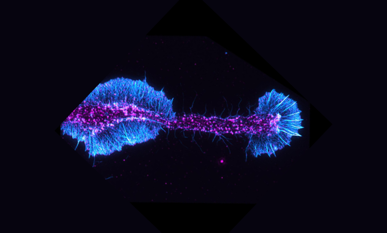
Banner image at top of page shows actin filaments (cyan) and an actin regulatory protein (magenta) in a differentiating oligodendrocyte. Image Credit: Brad Zuchero and Andrew Olson. Image acquired at the Neurosciences Microscopy Service, a Community Laboratory of the Wu Tsai Neurosciences Institute. The authors also acknowledge research support from the Gene Vector and Virus Core, another Wu Tsai Neuro community laboratory.