US triathlon star is ignoring ‘armchair quarterbacks’ as he plots IRONMAN Pro Series bid
“Having to get into a sprint finish for the last paycheck 50 weeks into the season did not feel good.”
That was how American star Matt Hanson, one of long-distance triathlon’s best runners, signed off his 2024 campaign, as his final kick at the IRONMAN 70.3 World Championships in New Zealand rounded off a rollercoaster IRONMAN Pro Series for him.
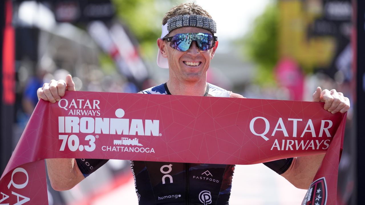
He’d been the long-time leader in the battle for the $200,000 first prize thanks to top 10 finishes at the IRONMAN World Championship, IRONMAN Texas and IRONMAN Lake Placid as well as a 70.3 victory at Chattanooga.
But despite racing from early April through to mid-December his 15th place in Taupo plus the strong late-season rallies of others would see him drop down to fifth place in the Pro Series standings, though that still brought a welcome bonus of $50,000.
Putting in the hard yards
However, having had a rest, it’s now all about regrouping and going again and he’s currently putting in the hard yards in training as his latest video, embedded below, shows.
He’s also busy plotting out his 2025 race season, saying: “I was thrilled to have finished fifth in the IRONMAN Pro Series last year.
“That wasn’t the end goal obviously, especially after leading it so long.
“But I’m chasing it again – hopefully we can equal it or better it. There’s a lot of new names coming to the list this year but I don’t think a lot of people would have picked me to be in the top 10 at the start of last year – they definitely wouldn’t have picked me to win a Pro Series 70.3 and we made it happen so I try not to let the armchair quarterbacks out there dictate the schedule too much.”
‘See where I’m stacking up’
Joining Hanson in focussing on the Pro Series this year will be Norwegian duo Kristian Blummenfelt and Gustav Iden, plus Magnus Ditlev, who has been third and second in the last two runnings of the IRONMAN World Championship.
And Hanson’s opening race of the season will see him tackle Denmark’s Ditlev at IRONMAN South Africa on 30 March.
He explained: “South Africa has been a bucket list race which I’ve wanted to do for a long time but it’s never really fitted in.
“It doesn’t really fit in this year either but I’ve got to force things a little bit earlier in the year, especially with racing so late into last year.
“But I think that’s going to be a good opportunity to go out and just see where everyone is at and see where I’m stacking up early in the year.
“A good chunk of the people chasing the Pro Series will go to South Africa and a good chunk will go to Oceanside [the following week].”
In terms of what comes after South Africa, he added: “Obviously IRONMAN Texas (26 April) has been on my list every year bar one as a pro and will be again and then I’ll do 70.3 St George (10 May).
“Then after that I’ll most likely to Eagleman, see how the points are shaking out and then reassess.”